Our missions:
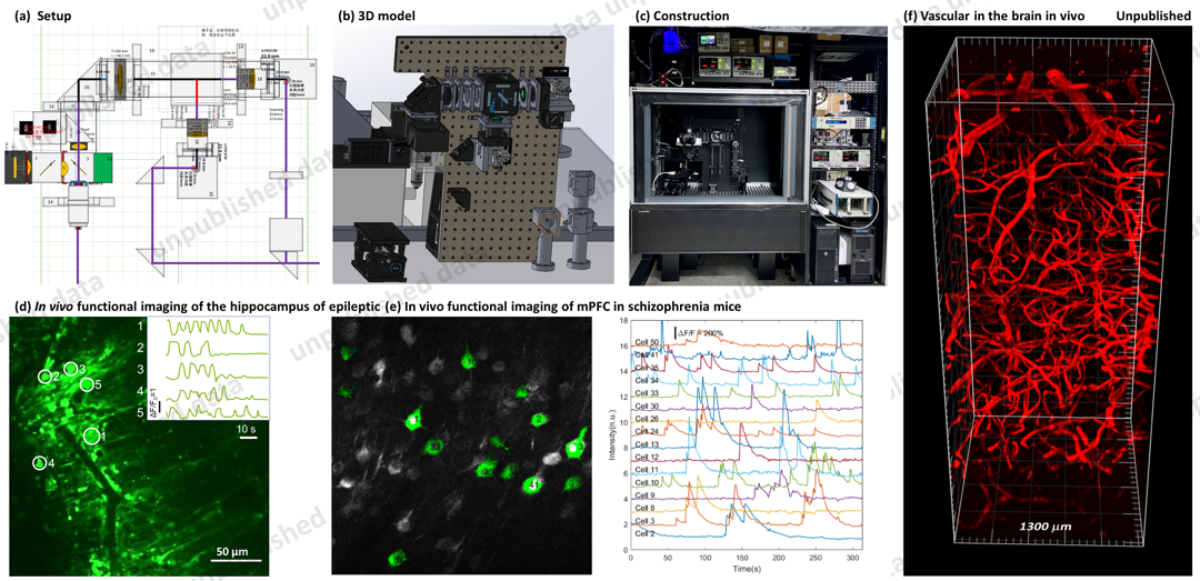
The Neural Imaging Lab is dedicated to the development of advanced microscopy technologies, such as multiphoton fluorescence microscopy and their application in investigating neural structure and function within the brain.
1. Deeper Penetration: Up to 2 mm
Three-photon imaging uniquely enables imaging of brain areas deeper than 1 mm in living animals. Our team is committed to enhancing this technique through advancements in laser technology, improvements in microscope capabilities, and refinement of image-processing algorithms. We aim to apply these innovations to study neural mechanisms in brain disorders involving deep brain regions like the hippocampus, striatum, and mPFC. Furthermore, three-photon imaging is expected to play a crucial role in the examination of organoids and marmoset brains.

2. Faster Speed: 1000 frames/s; Larger FOV: 6 mm x 6 mm
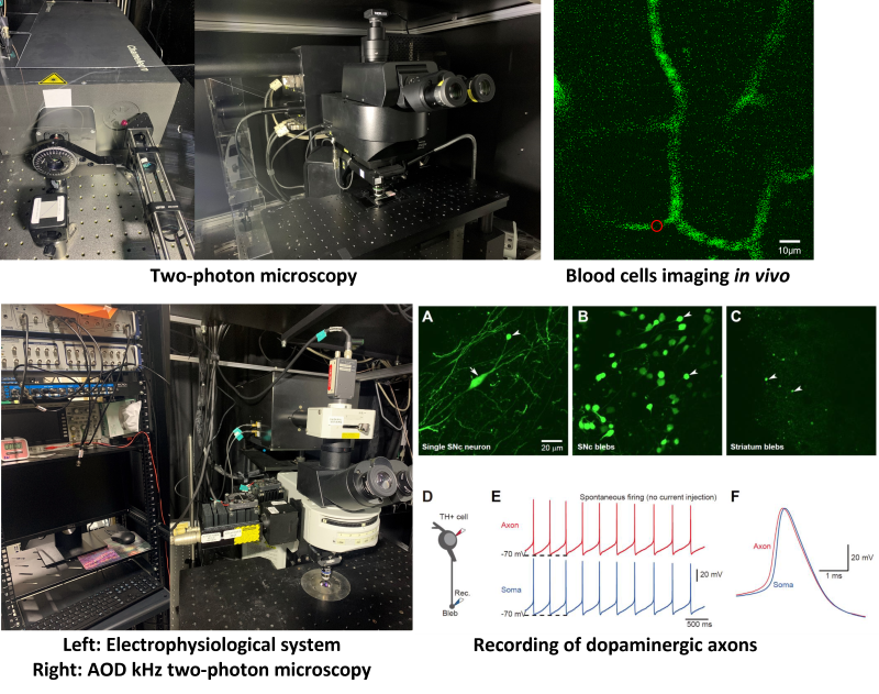
Two-photon imaging is a leading technology for in vivo neurofunctional imaging. Our group endeavors to make significant improvements in this technique by addressing two main challenges: 1) increasing the imaging speed from 30 Hz to over 1000 Hz for high-quality voltage fluorescence imaging, and 2) expanding the field of view from 0.5x0.5 mm to 6x6 mm to enable simultaneous recording of activities in multiple brain regions and analysis of their functional and structural connectivity.

3. Optical Stimulation of Neuronal Assemblies During Imaging
Optogenetics represents a groundbreaking technology in neuroscience. By combining two-photon excitation with optogenetics, we can simultaneously stimulate multiple individual neurons. Our unique approach concentrates on implementing three-dimensional light stimulation using advanced algorithms. In addition, we are developing a commercial two-photon optogenetic module, which can be easily integrated with existing commercial microscopes for simultaneous three-dimensional light stimulation and optical imaging.

